Email-ID: karanjulka1@gmail.com

Best gastroenterologist doctor in kalyan

                                             OGD SCOPY/UPPER GI ENDOSCOPY

Best gastroenterologist doctor in kalyan

UPPER GI ENDOSCOPY OR OGD SCOPY IS A MINIMALLY INVASIVE PROCEDURE TO INVESTIGATE AND TREAT PROBLEMS OF UPPER GASTROINTESTINAL TRACT. IT IS BOTH IDAGNOSTIC AND THERAPEUTIC PROCEDURE DONE UNDER LOCAL OR SEDATION. IT IS USED TO TREAT OESOPHAGEAL VARICES, STRICTURES, ULCER BLEEDS AND HIATAL HERNIA.

Upper GI endoscopy (sometimes called a ‘gastroscopy’ or simply an ‘endoscopy’) is a test which allows the doctor to assess the food pipe the stomach and around the first part of the small intestine – the duodenum. A small tube with camera is passed through your mouth into the stomach. Looking thru the tube, we get a clear view of the stomach. Sometimes the doctor takes sample of tissue for analysis in the laboratory.
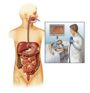
Best-gastroenterologist doctor-in-kalyan

Preparation before the procedure

You will be asked not to have anything to eat or drink for at least five to six hours before the test. When you come to the department, a doctor will explain the test to you and will usually ask you to sign a consent form. If you have any questions at this stage don’t be afraid to ask. It will also be necessary for you to remove any false teeth. They will be kept safely until after the examination.

During the test

In the examination room you will be made comfortable on a couch, resting on your left side. A nurse will stay with you throughout the test. Some doctors may spray a local anaesthetic on the back of your throat or give you a tablet to suck to numb the area. To keep your mouth slightly open, a mouthpiece will be put gently between your teeth. The procedure will be a painless one. During this time some air will be passed down the tube to distend the stomach and allow the doctor a clearer view.

After the test

You will be left to rest in the unit for at least thirty minutes. After this you can eat and drink normally. The back of your throat may feel sore for the rest of the day. You may also feel a little bloated if some of the air has remained in your stomach. Both these discomforts will pass, and need no medication.
                                      LOWER GI ENDOSCOPY / COLONOSCOPY

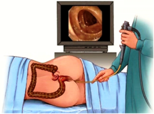
COLONOSCOPY OR LOWER GI ENDOSCOPY IS A MINIMALLY INVASIVE TECHNIQUE USED TO EVALUATE DISORDERS OF LOWER GASTROINTESTINAL TRACT. IT IS DONE UNDER SEDATION AND IS BOTH DIAGNOSTIC AND THERAPEUTIC PROCEDURE. THE ENTIRE LARGE INTERSINE IS EVALUATED UP TO TERMINAL ILEUM.

Colonoscopy is a test which allows the doctor to look at large intestine. In order to do the test a long tube with a camera is carefully passed through the anus into the large intestine. The video camera on the colonoscope transmits images of the inside of the colon to a monitor allowing the physician to assess for any abnormal findings.

Best-gastroenterologist-doctor-in-kalyan

Best-gastroenterologist-doctor-in-kalyan

The physician may take a sample of the lining of the bowel for closer examination under the microscope. A small piece of tissue is removed painlessly through the colonoscope, using tiny biopsy forceps.

It is also possible to remove adenomas (polyps) during colonoscopy. Polyps are abnormal projections of tissue, rather like a wart, which the doctor will want to remove and examine in more detail.

Preparation

To allow a clear view, the colon must be completely empty of waste material. Bowel preparation instructions would be given to you a day prior to the procedure.

It is important to take all of the laxative prescribed and also considerably increase your intake of clear fluids on the day before the examination, which will help clean the bowel.

When you come to the department, a member of staff will explain the test to you and will usually ask you to sign a consent form.

What will happen?

You will be placed in a comfortable position on your left side, and may be given medication by injection through a vein to make you sleepy and relaxed. You may experience some abdominal cramping and pressure from the air which is introduced into your colon. This is normal, and will pass quickly. You may also be asked to change position during the examination, and will be assisted by a nurse. The examination takes 15-60 minutes.

In many cases the doctor will be able to tell you the results of the test as soon as you are awake. However if a biopsy sample or polyp was removed for closer examination, these results may take up to ten days to process.

Many patients find that sedation tends to make them forget any explanations that may have been given them. Details of any results or further investigations may be obtained from your GP or specialist (whoever referred you for the test).

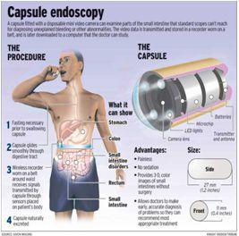
Capsule endoscopy is a procedure that uses a tiny wireless camera to take pictures of your digestive tract. A capsule endoscopy camera sits inside a vitamin-size capsule you swallow. As the capsule travels through your digestive tract, the camera takes thousands of pictures that are transmitted to a recorder you wear on a belt around your waist.

Capsule endoscopy helps doctors see inside your small intestine — an area that isn’t easily reached with more-traditional endoscopy procedures. Traditional endoscopy involves passing a long, flexible tube equipped with a video camera down your throat or through your rectum.

Capsule Endoscopy involves ingesting a small and safe capsule, which contains a  camera, capable of taking multiple images. The camera takes two pictures every second for eight hours, transmitting images to a data recorder about the size of a portable CD player that patients wear around the waist.

Best-gastroenterologist-doctor-in-kalyan

Best-gastroenterologist-doctor-in-kalyan

Capsule endoscopy assists in diagnosing gastrointestinal conditions such as gastrointestinal bleeding, malabsorption, chronic abdominal pain, and chronic loos emotions.
Once swallowed the camera moves naturally through the digestive tract while patients carry out their normal activities. Approximately eight hours after ingesting the camera, patients return the recording device to their doctor or nurse so the images can be downloaded to a computer and evaluated.

Instructions

Normal breakfast and lunchtime meal then clear fluids until 10.00pm
Nothing by mouth from 10.00pm until the start of the test

Procedure Day

Arrive in the unit between 8.00am and 8.30am
Admission and placement of sensor array and data recorder
Capsule swallowed with small amount of water
2 hours after ingestion of capsule clear fluids can be started
A light snack may then be eaten approximately 4 hours after swallowing the capsule
8 hours after the swallowing the capsule a normal diet may be resumed.

Endoscopic Retrograde Cholangio-Pancreatography

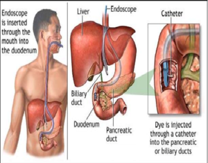
An ERCP (Endoscopic Retrograde Cholangio-Pancreatography) procedure allows the doctor to examine the bile duct and/or pancreas. During the procedure, a fine soft tube will be placed into one nostril to give you a little oxygen to breathe during the test. You will be given an injection into your blood which will make you very sleepy. Once you are sedated, an endoscope will be passed through your mouth, down into your stomach and the upper part of the small intestine. Contrast dye will be injected down the endoscope so that the pancreas and bile ducts may be seen on X-ray films. If the X-rays show a gallstone, the doctor will enlarge the opening of the bile duct. This is done with an electrically heated wire (diathermy) which you will not feel. Any stones will be collected into a tiny basket or left to pass into the intestine. If a narrowing is found, bile can be drained by leaving a short plastic tube stent in the bile duct. You will not be aware of the presence of the tube which will remain in place permanently. In certain cases, it may be necessary to replace the tube some months later if it becomes blocked.

Best-gastroenterologist-doctor-in-kalyan

Best-gastroenterologist-doctor-in-kalyan

Before the test

You will fasting for at least six hours before the procedure. When you come to the department, the procedure will be explained and a doctor will ask you to sign a consent form. It will also be necessary for you to remove any false teeth or contact lenses. Jewellery or metal objects should also be removed because they interfere with X-rays and a special instrument called aelectrocautery. They will be kept safely until after the examination.

After the test

When you return to the recovery room, you will feel sleepy. The nurse will advise you when you can eat and drink. You will be told the results of the test and what treatment has been given.

Going home

If you are having an ERCP without any additional treatment it is likely that you will be allowed home after the test, but it is essential that someone comes to collect you. Once home, it is important to rest quietly for the remainder of the day. Sedation lasts longer than you think.

What are the risks and complications?

Fortunately, these procedures are very safe – this is why they are used instead of surgery. If you have any problems after ERCP which you feel may be related to the test, please inform your doctor or hospital staff at once. An operation may be necessary to treat a complication, but this is very rare and can be managed well.

THIS IS A USEFUL PROCEDURE TO EVALUATED PATIENTS SUSPECTED OF HAVING FRESH BLOOD IN STOOLS. IT CAN HELP TO ACCURATELY DIAGNOSE THE CAUSE OF BLEED. IT IS DONE UNDER CONSCIOUS SEDATION.

Endoscopic Bariatric Interventions

Best-gastroenterologist-doctor-in-kalyan

WITH THE LATEST ADVENT OF ENDOSCOPIC BARIATRIC INTERVENTIONS, THE TREATMENT OF OBESITY HAS EVOLVED FROM SURGERY TO ENDOSCOPY. IT EMPLOYS INSERTION OF INTRAGASTRIC BALLOONS, ENDOSCOPIC SLEEVE GASTROPLASTY. THESE ARE NOT ONLY HELPFUL FOR WEIGHT REDUCTION BUT ALSO FOR TREATMENT OF NASH, SLEEP APNOEA.THE BIGGEST ADVANTAGE IS THAT IT IS A REVERSIBLE PROCEDURE.

Endoscopic bariatric interventions or endoscopic and metabolic therapies (EMBTs) are minimally invasive procedures performed to modify the structure and function of the gastrointestinal system, thereby facilitating to control metabolic diseases, such as diabetes, obesity and diabesity.

For Obesity

Following are the endoscopic procedures performed to control and treat obesity.

Intragastric Balloon

Intragastric balloons, as the name suggests, are balloon-like devices, made usually of silicon, placed inside the stomach

A patient may undergo endoscopic intragastric balloon therapy if he or she has a body mass index (BMI) between 30 and 40, is agreeing to commit to permanent lifestyle changes, and has no previous stomach or esophageal surgery.

Intragastric balloon is usually very safe procedure and  done as an outpatient procedure, and require minimal anesthesia. During the procedure, an endoscope  loaded with the balloon device is inserted through the mouth and guided to the stomach. The balloon device is placed in the stomach and inflated in position, and the endoscope is removed.

Endoscopic Sleeve Gastroplasty (ESG)

Endoscopic sleeve gastroplasty is a minimally invasive technique that facilitates weight loss. In this technique, an endoscope is inserted into the stomach through the mouth. The stomach is sutured along the outer curve to fold itself leading to a banana-sized tube. The stomach size is reduced by 70% approximately. This promotes early satiety and reduces food intake.

Endoscopic sleeve gastroplasty may be done in patients who have a body mass index (BMI) above 30, have failed in losing weight despite lifestyle modification, and do not wish to undergo traditional bariatric surgeries.

The procedure can be performed as an outpatient procedure, with minimal anesthesia, and the patient can go home the same day. However, the patient needs to commit to dietary modifications and regular follow-ups for the best outcome.

THESE ARE PROCEDURES INVOLVING APPLICATION OF BANDS TO BLEEDING PILES. IT IS CONSIDERED AS A PAINLESS PROCEDURE AND PATIENT IS DISCHARGED THE VERY SAME DAY.

WITH THE ADVANCEMENTS IN TECHNIQUES OF ENDOSCOPY, REMOVAL OF POLYPS HAVE BECOME A ROUTINE PROCEDURE. IT IS A VERY USEFUL TECHNIQUE FOR AVOIDING SURGICAL INTERVENTIONS FOR EARLY TUMORS.

WITH THE ADVENT OF A VARIETY OF SELF EXPANDABLE STENTS AND BALLOONS, THE TREATMENT OF STRICTURES HAS EVOLVED AND SHOWING ENCOURAGING RESLUTS. WE VERY RARELY NOWDAYS REQUIRE SURGICAL INTERVENTION FOR THEIR MANAGEMENT.

THIS DIFFICULT CONDITION HAS BEEN VERY SUCCESSFULLY BEING TREATED WITH POEMS VIA ENDOSOCPY. WE NO LONGER REQUIRE THE PATIENTS TO UNDERGO PAINFULL BALLOON DILATIONS FOR IT.

Best-gastroenterologist-doctor-in-kalyan

A VARIETY OF PLASTIC AND METAL STENTS ARE USED TO TREAT MALIGNANCIES AND STRICTURES OF GI SYSTEM.THIS SAVES THE PATIENT AN UNNECESSARY SURGERY FOR ADVANCED TUMORS AND IS A DAYCARE PROCEDURE.

Endoscopic Ultrasound (EUS)

Best-gastroenterologist-doctor-in-kalyan

Endoscopic ultrasound (EUS) is a specialized endoscopy procedure, where a specialize tube with an inbuilt small ultrasound device, is passed inside through the mouth into the upper part of the stomach. EUS offers unique advantage of precise and deeper visualization of surrounding peri-luminal organs and structures and also layers of the wall of GIT e.g. pancreas, bile duct, gallbladder, adrenal, and structures located deeper parts of chest (mediastinum) and abdomen.

EUS is generally an outpatient procedure done performed after 6 to 8 hours of fasting. EUS is a very safe procedure done under sedation that is monitored by an anaesthetist. The average ‘diagnostic EUS’ lasts about 20-30 minutes, and the patient can go home after 2-4 hours of observation. The ‘interventional EUS’ may require between 30-60 minutes to complete and is majorly performed as an inpatient procedure.

The main indications for ‘EUS’ are:

  1. Evaluation of various pancreas disorders –
  2. Bile duct disease.
  3. GIT sub-epithelial abnormalities e.g. polyp, leiomyoma, lipoma, GIST.
  4. Accurate cancer staging of oesophagus, stomach, pancreas, bile duct, ampulla.
  5. Mediastinal disease e.g. Lymph nodes, Lung cancer.
  6. Obtaining biopsy or cancer cells for diagnosing suspected tumours by EUS guided FNA.

The main indications for ‘interventional EUS’ are:

  1. Drainage internally of pancreatic fluid collections (
  2. Biliary drainage internally.
  3. Celiac plexus block for abdominal pain relief in patients with advanced pancreatic cancers.

Disease Spectrum

chronic Bloating of Stomach
obesity
Indigestion
Chronic Constipation or Diarrhea
Blood in Stools or Vomit
Anemia
Jaundice
Chronic Headaches With Acidity
Alcoholism
Loss of Appetite and Weight Loss
Black Colored Stools
KALYAN GASTRO CENTER
close slider

KALYAN GASTRO CENTER

Address: 1st Floor, Balaji Paradise bldg,

Near Commissioner Bunglow, Syndicate,

Santoshi Mata Road, Kalyan West. Pin 421301

Contact No : 70452 71331 / 70452 73517

DAILY 10 am To 3 pm, EXCEPT SUNDAYS